



When a child is diagnosed with cancer, parents are usually loaded with questions like, “Where is the tumour? Has it spread? How active is the disease? And is the current treatment working?”
Regular scans like CT and MRI can provide important structural information, such as the size and location of a tumour and how much space it is occupying. But sometimes, a scan cannot clearly tell whether a lump that remains after treatment contains active cancer or is simply scar tissue or another treatment-related change.
You also have to understand that cancer is not only a change in the body's structure. It changes the biological and chemical processes inside cells. Cancer cells divide and multiply rapidly, and many types of cancer cells consume nutrients such as glucose at a higher rate than normal cells.
This is where nuclear medicine offers a different way of looking at cancer. It can help doctors study certain biological activities inside the body and, in selected cancers, use those same biological targets to guide treatment.
So, can nuclear medicine change the way childhood cancers are diagnosed, monitored and treated? To understand that, it helps to first understand what nuclear medicine actually does.
What Is Nuclear Medicine in Cancer Care?
Nuclear medicine works a little differently from a regular scan. Instead of only taking a picture of the body's structure, it gives doctors a way to follow what cells are doing.
So, what actually happens during a nuclear medicine scan?
- First, a small amount of a radiotracer is given through a vein, usually in the arm or hand. The tracer carries a radioactive signal that a special scanner can detect.
- The tracer is designed to behave like, or attach to, something specific in the body. For example, in an FDG PET scan, the tracer behaves much like glucose, which cells use for energy.
- Why glucose? Cancer cells need energy to keep growing and multiplying. Many cancer cells therefore take up more glucose than normal cells.
- The cancer cells take up FDG as if it were glucose. But FDG is not ordinary sugar. Once inside the cell, it cannot be processed in the same way, so it remains trapped there long enough to be detected.
- The PET scanner then detects the radioactive signal coming from the tracer. Areas that take up more FDG can appear brighter on the scan, showing increased metabolic activity.
This gives doctors information that a structural scan alone may not provide. A CT or MRI can show where a tumour is and what it looks like. Nuclear medicine can add another question: what is happening inside that tissue?
And FDG is only one example. Different radiotracers can follow different biological processes or target different molecules, which is what makes nuclear medicine useful for different diseases and clinical questions.
How Is Nuclear Medicine Different From CT and MRI?
The biggest advantage of nuclear medicine is that it can give doctors information about what is happening inside abnormal tissue, not just what that tissue looks like.
Consider a child who has completed cancer treatment. A follow-up CT or MRI may still show a lump where the tumour was. But that lump does not necessarily mean that active cancer remains.
It could be:
- Living cancer cells that are still active
- Scar tissue left behind after treatment
- Inflammation or other changes caused by the treatment itself
This is where nuclear medicine adds something important.
A PET scan uses a radiotracer such as FDG to look for areas with increased metabolic activity. If the remaining tissue takes up a significant amount of the tracer, it tells doctors that the cells in that area are biologically active. That can provide an important clue about whether the abnormality needs closer evaluation.
The scan does not simply say, "There is a lump here." It adds another layer of information:
"There is a lump here, and this is how active the tissue appears to be."
When PET is combined with CT, doctors can then match that biological activity with its exact location in the body.
So, nuclear medicine does not compete with CT or MRI. It adds information that they may not provide on their own. That extra information can help doctors make a more informed decision about what the abnormal tissue represents and what should happen next.
How Can Nuclear Medicine Help Children With Cancer?
Nuclear medicine is not used in exactly the same way for every childhood cancer. The tracer doctors choose depends on what they are trying to find in the tumour and how that cancer behaves.
That is what makes the field particularly interesting. In some cancers, nuclear medicine helps doctors find and measure active disease. In others, it can provide information that conventional imaging may not answer clearly. And in selected cancers, the same biological targeting can even be used to deliver treatment.
Here are some of the areas where nuclear medicine is already making a difference.
Benefits of Nuclear Medicine in Childhood Cancer Care
Brain Tumours: Looking Beyond What an MRI Can Show
Brain tumours are the second most common childhood cancers after leukaemia, and to detect this, MRI remains the main imaging test. But sometimes an MRI cannot clearly tell whether a suspicious area is an active tumour, a treatment-related change or recurrent disease.
This is where specialised nuclear medicine tracers can add another layer of information.
FDG, the glucose-based tracer we discussed earlier, has limitations in the brain because healthy brain tissue naturally uses a lot of glucose. Newer amino-acid PET tracers, such as Methionine, FET and DOPA, can create better contrast between certain brain tumours and normal brain tissue.
This can help doctors:
- Detect tumour tissue more clearly
- Understand the extent of a tumour
- Identify biologically active areas for a biopsy
- Plan surgery more precisely
- Distinguish possible recurrence from treatment-related changes
So, in brain tumours, nuclear medicine is not trying to replace MRI. It can answer questions that MRI may leave behind.
Lymphoma: Finding Active Cancer, Not Just Enlarged Lymph Nodes
Lymphoma gives us another good example.
A lymph node can become enlarged for several reasons. A conventional scan can show that the node is bigger, but the more important question is whether it contains active lymphoma.
FDG PET-CT can help answer that question by showing areas where the cancer is taking up more glucose-like tracer.
This makes PET-CT particularly useful in many childhood Hodgkin and non-Hodgkin lymphomas for staging and assessing treatment response. Doctors can look not only at whether lymph nodes have changed in size, but also at whether their metabolic activity has reduced after treatment.
Neuroblastoma: When Nuclear Medicine Can Find and Treat the Cancer
First, what is neuroblastoma?
Neuroblastoma is a cancer that develops from immature nerve cells. It mainly affects young children and can develop in different parts of the body, including the abdomen, chest and areas near the spine.
Many neuroblastoma cells have a particular feature: they take up a substance called MIBG.
This gives nuclear medicine specialists something very useful to work with.
So, how can MIBG help doctors find neuroblastoma?
Scientists can make a radioactive version of the MIBG molecule. It behaves like MIBG inside the body, so neuroblastoma cells take it up.
Think of it as giving the cancer cells something they recognise and naturally collect.
The radioactive part does something the ordinary MIBG molecule cannot do: it gives off a signal that a nuclear medicine scanner can detect.
Once the radioactive MIBG reaches the neuroblastoma cells, the scanner can detect where the signal is coming from and create an image.
This can help doctors:
- Find neuroblastoma cells in different parts of the body
- Understand how far the disease has spread
- Measure the amount of disease
- Monitor how the cancer responds to treatment
So here, nuclear medicine is using the behaviour of the cancer cells themselves to find them.
But what if we use the same idea to treat the cancer?
This is where things become particularly interesting.
Instead of using radioactive MIBG only in a small amount to create an image, doctors can use a therapeutic radioactive form called I-131 MIBG in selected children.
The MIBG molecule still acts as the carrier. The neuroblastoma cells take it up because they recognise MIBG.
But this time, the radioactive iodine attached to the molecule delivers a much stronger dose of radiation around the targeted cancer cells.
In simple terms: Radioactive MIBG for imaging helps doctors find the cancer, and I-131 MIBG for treatment carries radiation towards the cancer cells.
This is called targeted radionuclide therapy. It does not mean the radiation affects only cancer cells, which is why doctors carefully select patients, assess the tumour's MIBG uptake and plan the treatment dose.
Is this what doctors mean by Theranostics?
This idea of using one biological target for both finding and treating disease is called theranostics.
Neuroblastoma gives us a good example:
Find the target → confirm that the cancer takes it up → use the same target to deliver treatment.
That is what makes nuclear medicine particularly exciting. It is not limited to showing doctors where a disease is. In selected cancers, it can use the disease's own biology to help guide treatment towards it.
What Are the Limitations of Nuclear Medicine in Childhood Cancer?
Nuclear medicine can add valuable information to cancer care, but it does not replace every other test or work for every type of cancer.
- It is not suitable for every cancer: Different cancers require different tracers and biological targets.
- Other tests may still be needed: CT, MRI, biopsy and blood tests can remain important for diagnosis and treatment planning.
- It requires careful interpretation: Tracer uptake can sometimes occur with inflammation or infection, so doctors consider the child's complete clinical picture.
- Targeted treatments are selective: Therapies such as I-131 MIBG are used only when the cancer has the right biological target and the child is suitable for treatment.
Nuclear Medicine and Molecular Theranostics at Sarvodaya Hospital
At Sarvodaya Cancer Institute, nuclear medicine combines advanced molecular imaging with targeted radionuclide therapies for selected cancers.
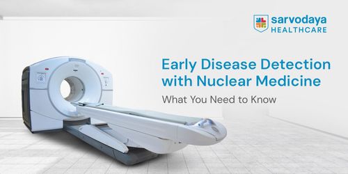
- Advanced imaging: The centre offers technologies such as 4D PET-CT and Gamma Camera imaging for diagnosis, staging and treatment assessment.
- Targeted therapies: Treatment options include I-131 MIBG therapy, PRRT, PSMA-based therapies, radioactive iodine therapy and microsphere therapy, depending on the cancer and its biological characteristics.
- Childhood cancer care: I-131 MIBG therapy is available for appropriately selected children with neuroblastoma whose tumours show the required MIBG uptake.
- Theranostics: The centre combines molecular imaging with targeted treatment, helping specialists identify suitable biological targets and use them to guide therapy.
- Specialist and multidisciplinary care: Nuclear medicine specialists work alongside oncology and other teams as part of the child's overall cancer-care plan.
- Recognised quality standards: The centre is EARL-certified by the European Association of Nuclear Medicine for standardised PET imaging.