
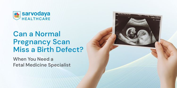


Pregnancy scans have transformed modern prenatal care. Today, doctors can monitor a baby's growth, evaluate developing organs, identify many structural abnormalities, and assess the overall health of a pregnancy long before birth. For most families, these routine scans provide reassurance that the baby is developing as expected.
Yet prenatal diagnosis remains one of the most challenging areas of medicine.
According to estimates, more than 1.7 million babies are born with birth defects every year in India, making congenital anomalies one of the leading causes of neonatal illness, infant mortality, and long-term disability. While many birth defects can be identified during pregnancy, others become apparent only at birth or even months later. This naturally raises an important question: If pregnancy scans are performed regularly, why aren't all birth defects detected before birth?
The answer lies in understanding how a baby develops inside the womb, what birth defects actually are, and what each pregnancy scan is designed to evaluate. Once these concepts become clear, the role of a fetal medicine specialist also becomes much easier to understand.
What Are Birth Defects?
The term birth defect does not refer to a single disease. It describes a broad group of conditions that affect how a baby develops before birth. These conditions may alter the structure of an organ, affect how it functions, or influence the baby's physical and neurological development.
Birth defects can involve almost any part of the body, including:
- Brain and nervous system: Spina bifida, anencephaly and hydrocephalus.
- Heart: Congenital heart defects affecting the chambers, valves or major blood vessels.
- Face and mouth: Cleft lip, cleft palate and other facial abnormalities.
- Spine, bones and limbs: Limb length differences, clubfoot or skeletal disorders.
- Kidneys and urinary tract: Missing, blocked or abnormally developed kidneys.
- Digestive system: Conditions affecting the oesophagus, intestines or abdominal wall.
- Chromosomes and genes: Conditions such as Down syndrome and several inherited genetic disorders.
- Body functions: Certain hearing, metabolic and neurological disorders that may not produce visible structural changes before birth.
The severity also varies considerably. Some birth defects require only regular follow-up after delivery, while others need specialised treatment immediately after birth or, in selected cases, even before the baby is born.
Understanding this diversity is important because every birth defect behaves differently. Some produce obvious anatomical changes during pregnancy, whereas others remain hidden despite appearing completely normal on routine imaging.
When Are Birth Defects Usually Detected?
Since the term birth defect refers to a group of many conditions, they don't become clear at the same time. Some can be identified during pregnancy, while others only become noticeable after birth, when the baby starts functioning independently outside the womb.
Before Birth
Many birth defects can be detected during pregnancy because they cause visible changes in the baby's developing organs. During detailed ultrasound examinations, doctors may identify conditions such as spina bifida, major congenital heart defects, kidney abnormalities, abdominal wall defects, and severe skeletal disorders.
At Birth
Some birth defects are present throughout pregnancy but are easier to recognise after the baby is born. This may happen because the condition is too subtle to detect on a routine scan or because it becomes more apparent once the baby is examined directly. Examples include certain forms of cleft palate, mild limb deformities, intestinal abnormalities, and some congenital heart defects.
After Birth
Some conditions, such as hearing loss, vision problems, metabolic disorders, or certain developmental disorders, may not be apparent during pregnancy because they do not produce obvious structural changes. Similarly, some abnormalities, such as a small heart defect or even a cleft lip in an unfavourable fetal position, may occasionally go undetected on routine scans. As a result, these conditions may only be diagnosed after birth, or sometimes weeks, months, or even years later.
This explains an important principle of prenatal diagnosis: the timing of detection depends on the nature of the condition, not simply on the number of scans performed.
Why Do Birth Defects Occur?
Parents often wonder whether they did something that caused the condition.
In most pregnancies, it's not the genuine cause.
Birth defects develop for many different reasons, and in a large number of cases, doctors cannot identify a single definite cause.
Factors that may increase the risk include:
- Genetic or chromosomal changes, which may occur spontaneously or run in families.
- Family history of inherited disorders.
- Maternal infections during pregnancy.
- Poorly controlled diabetes or certain other medical conditions.
- Deficiency of important nutrients such as folic acid.
- Exposure to harmful substances, including alcohol, tobacco and certain medications.
- Increasing maternal age for some chromosomal abnormalities.
Often, birth defects result from a combination of genetic and environmental factors rather than one isolated event. This is why every pregnancy carries some background risk, even when there is no family history or known medical problem.
How Do Routine Pregnancy Scans Monitor a Baby's Development?
Early Pregnancy Scan (6-10 Weeks)
The first ultrasound is performed to confirm that the pregnancy is developing normally. At this stage, the embryo is only a few millimetres long, so the focus is not on identifying birth defects but on establishing the basics of a healthy pregnancy. The scan confirms the baby's heartbeat, determines the number of babies, checks that the pregnancy is developing inside the uterus, and accurately estimates the gestational age and expected due date.
First Trimester Scan (11-14 Weeks)
As the baby grows, the first trimester scan provides the earliest opportunity to assess developing anatomy. Along with measuring the nuchal translucency (NT), doctors evaluate early structures such as the brain, limbs, stomach and bladder while screening for markers associated with certain chromosomal conditions, including Down syndrome. Although the baby's organs are still developing, this scan can identify several major abnormalities at an early stage and helps determine whether further evaluation is required.
Detailed Anomaly (Level II) Scan (18-22 Weeks)
By the middle of pregnancy, most major organs have developed sufficiently to allow a comprehensive anatomical examination. This is why the Level II or anomaly scan is considered the most important structural assessment during pregnancy. Doctors carefully examine the baby's brain, spine, face, heart, lungs, kidneys, abdomen and limbs, while also assessing the placenta, umbilical cord and amniotic fluid. Many significant structural birth defects can be detected during this examination, making it one of the most valuable milestones in prenatal care.
Growth and Wellbeing Scan (Third Trimester)
During the final months of pregnancy, the focus shifts from organ formation to the baby's growth and overall wellbeing. This scan monitors whether the baby is growing appropriately, evaluates the placenta, measures the amount of amniotic fluid and assesses blood flow using Doppler ultrasound. It also helps identify certain conditions that may become apparent later in pregnancy and provides important information for planning a safe delivery, particularly in high-risk pregnancies.
Why Can Routine Pregnancy Scans Miss Some Birth Defects?
Routine pregnancy scans are designed to monitor the baby's growth and detect many structural abnormalities. However, no ultrasound can identify every birth defect with complete accuracy. This does not indicate a limitation in medical care but reflects the nature of fetal development itself.
Some of the common reasons include:
- The baby's position: If the baby is facing the mother's spine or has its hands covering the face, certain abnormalities, such as cleft lip, may not be clearly visible.
- The stage of pregnancy: Some conditions develop gradually and may not become apparent until later in pregnancy.
- The size of the abnormality: Tiny defects, such as small openings between the chambers of the heart, can be difficult to detect on a routine scan.
- The type of condition: Some disorders affect how an organ functions rather than how it develops. Conditions involving hearing, metabolism, or certain genetic disorders may not produce visible changes on ultrasound.
- Technical limitations: Factors such as reduced amniotic fluid, multiple pregnancies, or maternal body habitus can sometimes affect image quality.
This is why, when a routine scan raises a concern or a pregnancy is considered high-risk, doctors may recommend a more detailed evaluation by a fetal medicine specialist.
What Does a Fetal Medicine Specialist Do Differently?
A fetal medicine specialist is an obstetrician with advanced training in diagnosing and managing fetal conditions before birth. Their role is not limited to performing another ultrasound. They help understand the nature of the finding, its possible cause, its impact on the pregnancy, and the best way to manage it.
A detailed fetal evaluation may include:
- Reviewing the complete pregnancy history, family history, and maternal health conditions.
- Performing specialised imaging such as Level II anomaly scans, fetal echocardiography, 3D/4D ultrasound, or Doppler studies whenever indicated.
- Assessing whether the finding represents a normal variation or a condition requiring treatment or closer monitoring.
- Recommending additional investigations, such as genetic counselling or diagnostic tests like amniocentesis, when clinically appropriate.
- Coordinating care with obstetricians, neonatologists, paediatric surgeons, genetic specialists, and other experts to plan pregnancy, delivery, and newborn care.
The goal is not simply to diagnose a condition, but to provide parents with the clearest possible information and prepare for the safest outcome for both mother and baby.
Can Birth Defects Be Prevented?
Not every birth defect can be prevented. Many result from genetic or chromosomal changes, while others occur without a clearly identifiable cause. However, certain measures can reduce the risk of some birth defects and support a healthier pregnancy.
Doctors generally recommend:
- Taking folic acid before conception and during early pregnancy to reduce the risk of neural tube defects.
- Managing medical conditions such as diabetes before and throughout pregnancy.
- Avoiding alcohol, smoking, recreational drugs, and unnecessary medications unless advised by your doctor.
- Discussing all prescription medicines and supplements with your healthcare provider before or during pregnancy.
- Staying up to date with recommended vaccinations and protecting yourself against infections during pregnancy.
- Maintaining a healthy weight before pregnancy and attending regular antenatal check-ups.
Even when a birth defect cannot be prevented, early diagnosis allows doctors to monitor the pregnancy closely, plan delivery appropriately, and arrange specialised newborn care whenever required. Early preparation can make a significant difference to pregnancy and neonatal outcomes.
Comprehensive Fetal Medicine Care at Sarvodaya Hospital
- Dedicated Centre for Fetal Medicine, providing specialised evaluation, diagnosis, monitoring, and management of both routine and high-risk pregnancies.
- Experienced fetal medicine specialists with expertise in managing fetal abnormalities, genetic concerns, multiple pregnancies, and other complex maternal-fetal conditions.
- Advanced prenatal imaging, including high-resolution ultrasound, Level II anomaly scans, 3D/4D ultrasound, fetal echocardiography, and Doppler studies for detailed fetal assessment.
- Pre-conception and antenatal counselling, helping couples understand potential risks, available screening options, and pregnancy planning before and during pregnancy.
- Specialised fetal interventions, where clinically appropriate, offering advanced in-utero procedures to improve fetal outcomes in selected conditions.
- Individualised pregnancy monitoring, with personalised care plans based on the baby's development, maternal health, and changing clinical needs.
- Multidisciplinary care under one roof, with seamless collaboration between fetal medicine specialists, obstetricians, neonatologists, paediatric surgeons, genetic counsellors, and other specialists.
- Advanced NICU and newborn support, ensuring continuity of care from prenatal diagnosis and delivery planning to specialised neonatal management whenever required.
- Compassionate emotional support, providing clear communication, counselling, and guidance to help families make informed decisions with confidence throughout their pregnancy.